SERVICES > DENTISTRY > Radiology Center

Radiology Center in Carabanchel

Our clinic is equipped with technology to perform all types of digital radiographs currently available on the market: Orthopantomography, cephalometric radiography, CBCT, intraoral, periapical, bitewing, and carpal (CARPO) X-rays.

We are your Radiology Center in Carabanchel.

Orthopantomography

Panoramic Radiograph

A panoramic radiograph or orthopantomogram is an extraoral X-ray that shows both jaws and the teeth. It is an image that provides a global view of the oral cavity and adjacent structures.

Panoramic radiography is a fundamental tool for the preliminary study of any dental treatment, and its use is very common in the initial patient assessment or for treatments such as oral surgery or orthodontics.

Orthopantomography is a very comfortable radiological test for the patient, as the level of radiation received is minimal, in addition to being painless and fast.

During the test, the patient must stand in front of the device, biting on a small piece, keeping the two arches of the mouth separated and the head upright.

In just a few seconds, the obtained image appears on the screen, and the dentist can analyze the test in detail.

At our Dental Clinic in Carabanchel, we have state-of-the-art digital technology that allows for precise orthopantomograms with the lowest radiation for the patient.

Radiological tests are delivered digitally upon request by the patient or another professional at our radiology center in Carabanchel.

Advanced Dental Technology Carabanchel
CBCT dental clinic Carabanchel

Cephalometric Radiography

Frontal or lateral extraoral radiography

Cephalometric radiography is an extraoral X-ray (frontal or lateral) performed at a greater distance than usual to prevent the image from being distorted by the structures of the skull.

In dentistry, lateral cephalometric radiographs of the skull are primarily used. These allow us to clearly see the facial bones and the patient’s teeth. They are part of orthodontic studies, as we use them to measure the angles and proportions of the patient’s facial bones, thus understanding the relationship between their facial structures through cephalometric analysis.

Thanks to the latest digital technology, we can carry out precise radiological studies quickly at our Dental Clinic in Carabanchel.

CBCT Scanner

Cone Beam Computed Tomography

The 3D CBCT scanner or Cone Beam Computed Tomography currently allows us to obtain an extremely precise three-dimensional (3D) image of the teeth, soft tissues, and bones of the oral cavity and adjacent areas of the patient.

At our Dental Clinic in Carabanchel, this radiological test is performed standing in an open machine that reduces the feeling of claustrophobia.

Through a single 360º rotation and in less than 30 seconds (with a much lower radiation dose than a conventional helical CT scan), we obtain a series of images that allow us to study the patient’s oral cavity in full detail.

Currently, it is used for dental implant procedures or oral surgery as well as for endodontics, orthodontics, and periodontics.

Oral Surgery Carabanchel

Intraoral Scanner

Patient comfort and precision

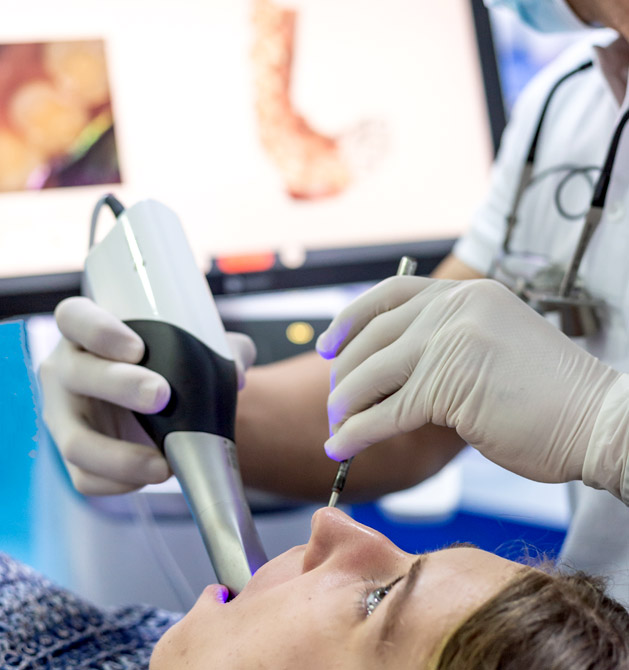
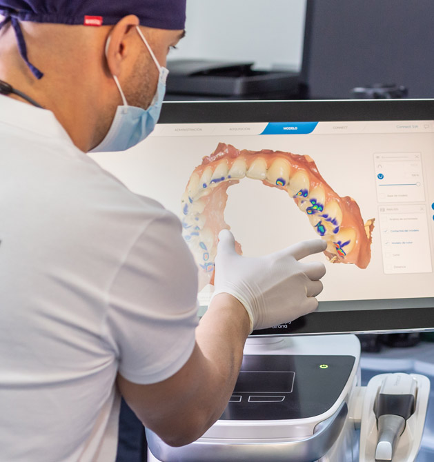
An intraoral scanner is a digital system that replaces or complements traditional impressions in dentistry.

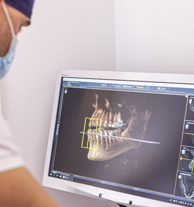
Using a small, easy-to-handle intraoral sensor, images of the patient’s mouth are obtained quickly and accurately, allowing us to reconstruct it three-dimensionally on a touch screen where we can analyze the digital model in detail. Subsequently, we can design, digitally print, or mill structures that allow us to offer greater precision in the fit.

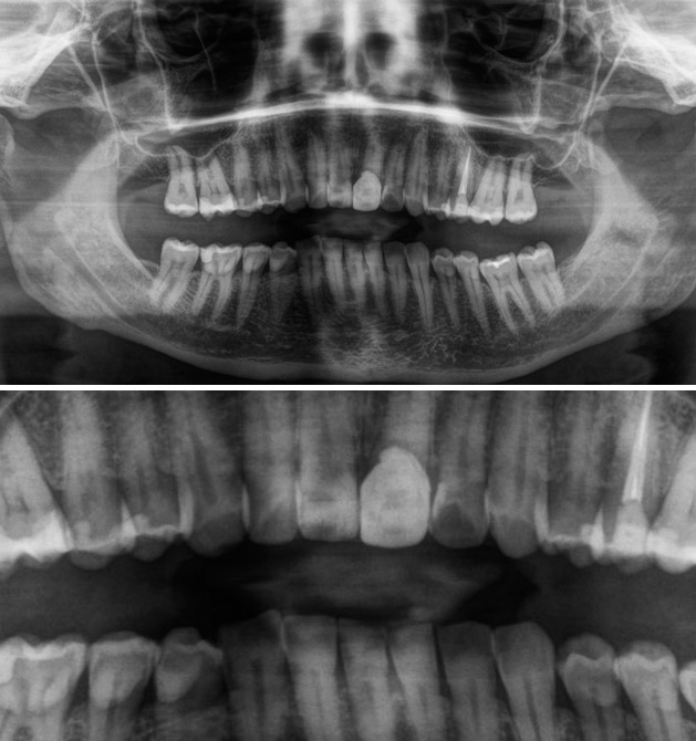
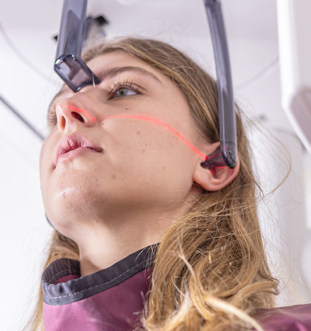
In dental implantology, it has many applications, as it facilitates, together with the CBCT scanner, the creation of surgical guides that help us position implant treatments previously planned digitally using powerful software.

Likewise, the preparation of prostheses on implants represents a leap in both quality and speed of treatments, as the connection and sending of the work is carried out through digital platforms almost instantaneously.

In orthodontics, it allows us to carry out a preliminary study as well as subsequent digital modifications until the final result, which makes it easier to explain to the patient both what the result of their treatment will be and the movements that will be necessary to achieve it. In this way, we have much more precise treatments and more controlled movements.

The instant connection to the cloud of the main aligner and invisible orthodontic systems facilitates the sending of information in less than 1 hour, providing significant agility.

The intraoral scanner is one of the most useful tools in the daily life of a clinic, as it allows us to capture images for study models and treatment planning for aesthetic dentistry, restorative dentistry, prosthetics, splints, and more.

Periapical Radiographs and Bitewings

Minimal radiation for the study of teeth and adjacent bone

These are intraoral radiographs that, with minimal radiation, provide great detail of the teeth and adjacent bone.

They are normally performed to accurately diagnose cavities, periodontal disease, or tooth fractures, among other pathologies.

Thanks to digital technology, these images are processed in a matter of seconds and provide much more clarity and precision than conventional ones.

CARPO

Specific study to determine bone age

During the orthodontic treatment of malocclusions, the evaluation of the patient’s bone growth is fundamental, given that most are at a developmental age.

Chronological age does not always coincide with bone maturity; therefore, the orthodontist sometimes requests a specific study to analyze what stage the patient’s bones are in and ensure that the orthodontic treatment modifies growth accordingly.

The most widespread method for determining a patient’s bone age is through a radiograph of the left hand, including the wrist. The parameters it offers are universally considered a true biological clock.

At our Dental Clinic in Carabanchel, we have the latest state-of-the-art digital technology in dental radiology, which allows us to obtain extremely precise images with the lowest radiation for the patient.

Privacy Overview

This website uses cookies so that we can provide you with the best user experience possible. Cookie information is stored in your browser and performs functions such as recognising you when you return to our website and helping our team to understand which sections of the website you find most interesting and useful.